CytoSMART – Live-Cell Imaging Microscopes – Omni
Being able to monitor cell cultures over time provides a great insight into their physiology and function. Live-cell imaging microscopes open up novel and exciting avenues to study cellular health, viability, colony formation, migration, and cellular responses to external stimuli.
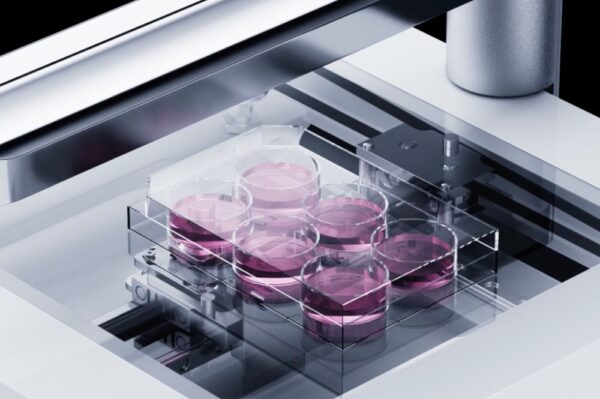
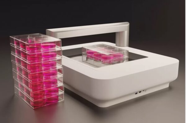
To help life science researchers improve their understanding of cellular processes, CytoSMART Technologies has developed an automated brightfield microscope that can visualize an entire surface of a cell culture vessel and operate from inside a standard CO2-incubator, biological safety cabinet, or on a benchtop. Not limited to a specific type or quantity of culture vessels, the CytoSMART Omni captures cellular behavior by creating high-quality time-lapse videos for days or even weeks at a time.
Applications
- Clonogenic assay
- Wound healing assay
- Cell confluence assessment
Technical Specifications
| Dimensions (L × W × H) | 396 mm × 345 mm × 171 mm |
| Weight | 9 kg |
|---|---|
| Optics | Brightfield with digital phase contrast |
| Magnification | 10× fixed objective |
| Light source | LED |
| Camera | 6.4 MP CMOS |
| Scan area | 99 mm × 131 mm |
| Exported formats | JPG, XLSX & MP4 |
| Well plate types | 6 – 384 well plates |
| Culture flask types | Petri dishes, T25 – T225, triple flasks, and HYPERFlasks |
| Other labware | Anything transparent and lower than 55 mm |
| Operating environment | 5 – 40 °C, 20 – 95% humidity |
| Support | Via email and live chat |
Key Features
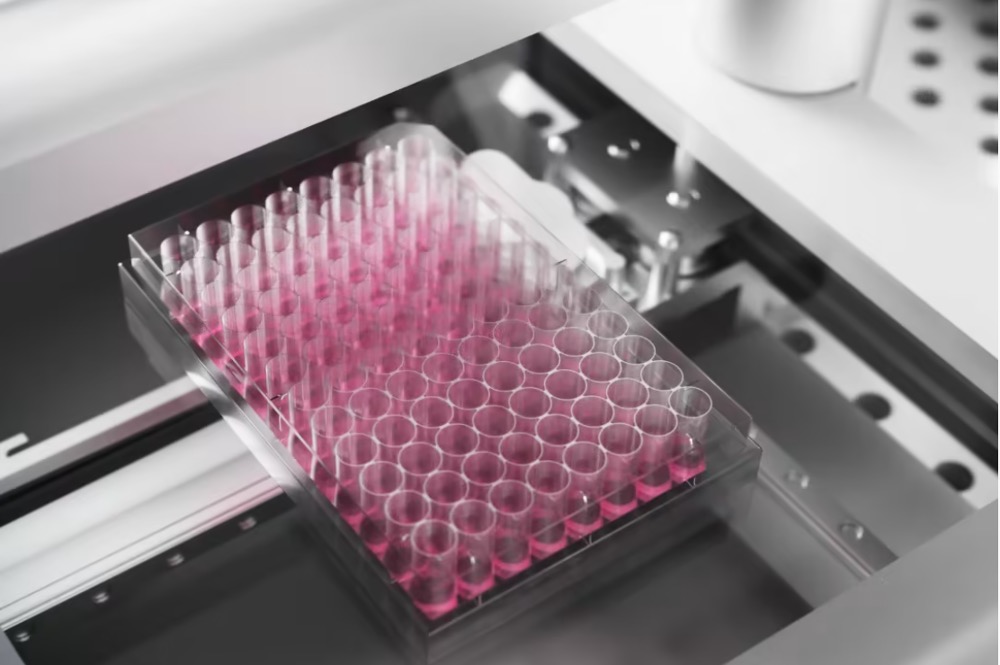
NEW – Automated Whole-Well Imaging With Higher Spatial Resolution
The CytoSMART Omni, fitted with the new digital 6.4 MP CMOS camera, can monitor cell cultures and study biological processes with enhanced spatial resolution, while also allowing cells to be kept in their desired culture environment. There is no need to move a culture vessel on the sample stage, since the camera moves along the platform to capture the entire area of the vessel. Higher quality images coupled with robust AI-based image analysis ensure that you will generate reliable and reproducible experimental data with minimal artifacts.
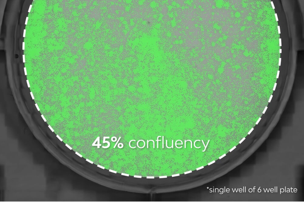
Complete Overview of Sample Confluency
Manual handling and cell seeding can cause a variable density distribution within culture vessels. Randomly selecting several areas of interest or tile scanning is a common practice to overcome that issue, however, this is either time-consuming to set up or to post-process. The CytoSMART Omni automatically scans complete well surface areas and instantly stitches these images to give users a complete overview of cell coverage.
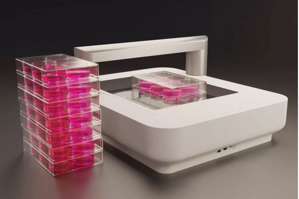
Efficient Screening of Culture Multiwell Plates
The CytoSMART Omni can scan up to 6 vessels per hour. High-throughput experiments are not limited to a single culture vessel. The CytoSMART Omni can process multiple plates in quick succession, automatically stitching acquired images and safely storing data for each individual plate.
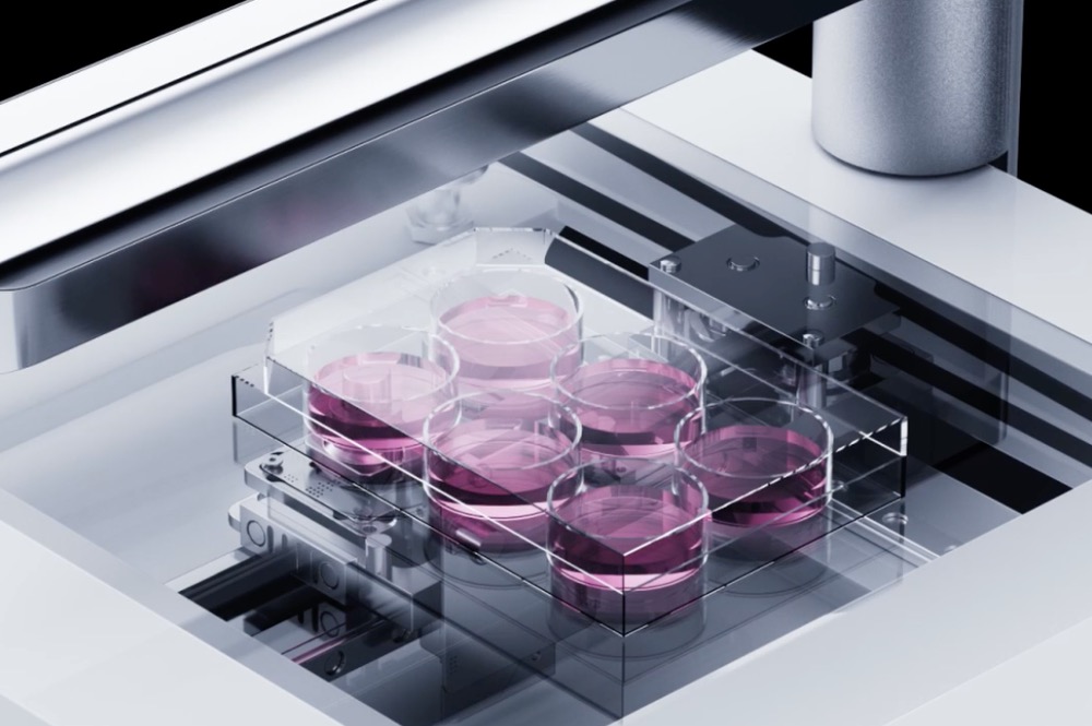
Compatibility With Any Transparent Culture Vessel
Cell-based assays and subculturing all benefit from standardization. Choosing the right vessel and going through the process of understanding how cells behave in the said vessel is vital for reproducibility. The CytoSMART Omni is highly flexible and compatible with any transparent vessel that is lower than 55 mm. All vessels with a surface area smaller than 99*131 mm can be imaged in full (6 – 384-well plates, T-25 – T225, Petri dishes, triple flasks, and HYPERFlasks).
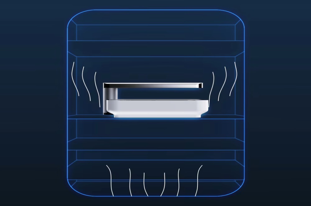
Perform Analysis at Desired Culturing Environment
The CytoSMART Omni is designed for your incubator. Live-cell imaging requires environmental control throughout the experiment. To maintain favorable cellular environment and minimize disturbance of the samples, users can place the compact CytoSMART Omni inside any standard cell culture incubator, where the imaging of cells is performed automatically via a moving camera that captures the entire area of a culture vessel.
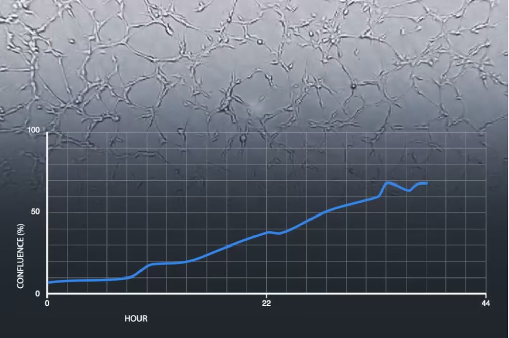
Continuous Imaging Provides Real-Time Insight into Cellular Processes
Perform kinetic assays over the course of days or even weeks. Time-lapse imaging allows you to pinpoint important events in the progression of the cell culture from experiment to experiment. Uncover attachment and detachment rates, evaluate events like cell death, and compare growth rates. Data collection using video monitoring allows users to capture changes within samples and compare the rate of change between samples.
CytoSMART™ Map View: Visualization and Data Analysis of Complete Cell Cultures
Wound Healing Test With CytoSMART OMNI Brightfield Live-Cell Imager
The CytoSMART Omni With Improved Optics
Key Industries
- Biopharmaceutical and Biotechnology
Similar Products

