Showing all 5 results

  • CytoSMART – Automated Fluorescence Microscopy – Lux3 FL

    The CytoSMART Lux3 FL is a small fluorescence live-cell imaging microscope equipped with one brightfield and two fluorescent channels (green and red). The device enables researchers to unravel cellular processes in real-time, pr...

    The CytoSMART Lux3 FL is a small fluorescence live-cell imaging microscope equipped with one brightfield and two fluorescent channels (green and red). The device enables researchers to unravel cellular processes in real-time, providing lots of valuable data about the experiments and answers if, when, how, and to what extend the cellular processes occur. CytoSMART’s first fluorescent cell imaging device allows users to track dynamic cellular processes with high specificity by taking high-quality images to create real-time time-lapse movies. Simultaneously, the cells are kept in a controlled environment inside a standard cell culture incubator.

    Let Lux3 FL capture fluorescent and brightfield images for you

    Currently, fluorescent labeling is mostly used as an end-point measurement. However, time-lapse imaging of live cells can give much more information about biological processes. By using automated imaging at regular time intervals, the temporal resolution of the fluorescent data is increased, leading to even more relevant data about the cellular processes. In this way, with fluorescence microscopy, researchers can not only determine if a certain process has occurred, but also when it occurred and at what speed.

  • CytoSMART – Compact Inverted Lab Microscope – Lux2

    The CytoSMART Lux2 provides an easy and cost-effective solution for time-lapse imaging. Monitor cell proliferation, motility and morphology for hours up to weeks at a time.

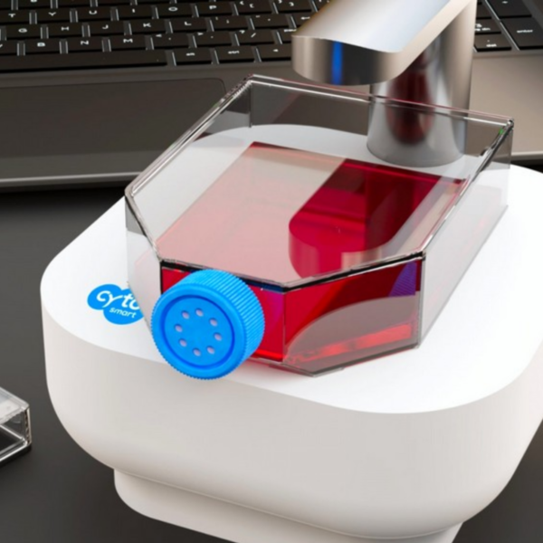
    The CytoSMART Lux2 is a compact inverted lab microscope for bright-...

    The CytoSMART Lux2 provides an easy and cost-effective solution for time-lapse imaging. Monitor cell proliferation, motility and morphology for hours up to weeks at a time.

    The CytoSMART Lux2 is a compact inverted lab microscope for bright-field live cell imaging. It works from inside any standard CO2-incubator and can be used in routine cell culture processes.

  • CytoSMART – Fluorescence Cell Counter – Exact FL

    The CytoSMART Exact FL is an automated, dual fluorescence cell counter with an expanded field of view and unmatched resolution and magnification power.

    Using an advanced CMOS-based optical system and an...

    The CytoSMART Exact FL is an automated, dual fluorescence cell counter with an expanded field of view and unmatched resolution and magnification power.

    Using an advanced CMOS-based optical system and an image analysis software powered by Artificial Intelligence (AI), the CytoSMART Exact FL calculates the exact number of cells in a sample and provides reliable assessment of key cellular parameters, including cell viability and transfection efficiency. The increased magnification and high resolution offer an unprecedented level of detail, allowing to distinguish between whole cells and cell debris and count even the smallest of mammalian cells (CAR-T cells, stem cells, PBMCs or splenocytes).

    Key Features

    • Multicount — increase counting accuracy by taking multiple images of one sample.
    • Robust declustering algorithm – identify individual cells in clusters.
    • Innovative organoid counting software – count organoids of varying shape and size.
    • Secure cloud data storage – safely store and access your experimental data.
    • Automates software updates – stay up-to-date with the latest software features.
    • Compatible with a range of cell counting slides – use your own counting chamber.
  • CytoSMART – Live-Cell Imaging Microscopes – Lux3 BR

    Live-cell imaging allows researchers to determine not only whether, but also how and when certain biological events occur in cell culture. In order to achieve the high image quality standards required for data analysis and publications, live-cell ...

    Live-cell imaging allows researchers to determine not only whether, but also how and when certain biological events occur in cell culture. In order to achieve the high image quality standards required for data analysis and publications, live-cell imaging is performed predominantly using a microscope with a stage top incubation box. However, the culture conditions in the incubation box are more prone to fluctuations, as compared to a dedicated cell culture incubator. Using the CytoSMART Lux3 BR, it is no longer necessary to choose between optimal cell culture conditions and image quality.

    Live-cell imaging has become increasingly valuable in the fields of cancer research, drug discovery, immunology, tissue engineering, stem cell research, and 3D cell culture models. Various experimental applications, including cell viability and cell differentiation monitoring, spheroid and organoid characterization, microfluidic chip platforms, cell morphology analysis, single-cell tracking, and angiogenesis monitoring can benefit from detailed kinetic visualization.

  • CytoSMART – Live-Cell Imaging Microscopes – Omni

    Being able to monitor cell cultures over time provides a great insight into their physiology and function. Live-cell imaging microscopes open up novel and exciting avenues to study cellular health, viability, colony formation, m...

    Being able to monitor cell cultures over time provides a great insight into their physiology and function. Live-cell imaging microscopes open up novel and exciting avenues to study cellular health, viability, colony formation, migration, and cellular responses to external stimuli.

    To help life science researchers improve their understanding of cellular processes, CytoSMART Technologies has developed an automated brightfield microscope that can visualize an entire surface of a cell culture vessel and operate from inside a standard CO2-incubator, biological safety cabinet, or on a benchtop. Not limited to a specific type or quantity of culture vessels, the CytoSMART Omni captures cellular behavior by creating high-quality time-lapse videos for days or even weeks at a time.

    Applications

    • Clonogenic assay
    • Wound healing assay
    • Cell confluence assessment