dSTORM
Direct Stochastic Optical Reconstruction Microscopy (dSTORM) relies on the stochastic activation of single fluorophores that blink from “off” to “on”, and quickly back to “off”. The process is repeated many times, activating just one molecule within a diffraction-limited region at a time.
Best for: very high resolution, molecular distributions in fixed cells
PALM
Photoactivated Localization Microscopy (PALM) uses photoactivatable fluorophores to resolve spatial details of tightly packed molecules. Once activated, fluorophores emit for a short period but eventually bleach. The laser stochastically activates fluorophores until all have emitted.
Best for: protein dynamics, organization and quantification, in fixed and live samples
PAINT
In Point Accumulation for Imaging in Nanoscale Topography (PAINT), single-molecule localizations are achieved using transient binding of fluorophores to targeted proteins. PAINT is commonly performed with DNA strands < 10 nt. Like other SMLM techniques, it can reach spatial resolutions of 20 nm.
Best for: biomolecular organization and quantification, in fixed samples
Single-particle tracking
Single-Particle Tracking (SPT) allows the motion of individual particles to be followed in vitro or in living cells, to obtain information on their dynamic behavior over time.Trajectories can be extracted with a resolution of up to milliseconds.
Best for: protein dynamics and tracking, in live samples or in solution
Single – molecule FRET (smFRET)
During single-molecule Förster Resonance Energy Transfer (smFRET), the emission energy of a donor fluorophore is transferred to an acceptor, which subsequently fluoresces when in close proximity. It enables distances between single molecules to be measured at a scale of 1-10 nanometers.
Best for very high resolution, molecular distributions in fixed cells
Technical Specifications
| Resolution | Single-molecule localization microscopy (SMLM) 20 nm in XY (incl. STORM, DNA-PAINT, PALM)* 3D: 100 nm in Z over a range of +/- 600 nm Widefield/TIRF Measured as FWHM of fluorescent signal (488 nm): 236 +/- 16 nm |
| Illumination Control | Option to select illumination angle: 0°-65° Epifluorescence/widefield | HILO | TIRF | LED array for bright-field imaging Flat-field homogeneous laser illumination: coefficient of variation <22% |
| Lasers | SMLM in multiple colors (4 lasers) and 2 simultaneous channels (dichroic mirror split at 640 nm) Power At sample: 405 nm (15 mW), 488 nm (250 mW), 561 nm (250 mW), 640 nm (250 mW) At source: 405 nm (1 W), 488 nm (1 W), 561 nm (750 mW), 640 nm (1 W) Switch on time 405 nm (1 ms), 488 nm (1 ms), 561 nm (3 ms), 640 nm (1 ms) |
| Power Density | 4 kW/cm2 (0.24 kW/cm2 for UV) Continuous laser |
| Temperature Control | Running temp 30-32°C Heating up to 42°C 1.5 – 2 hours warm up Sample temperature stable to +/- 0.1°C at steady state +/- 0.2°C with typical light program engaged** +/- 0.3°C with 100% laser power Precise temperature control, heating elements keep the entire unit at a uniform temperature. Ideal for live cell imaging and single-particle tracking |
| Product Dimensions | Nanoimager 21.5 cm (w) x 21.5 cm (d) x 15.5 cm (h) Light Engine 21.5 cm (w) x 42.0 cm (d) x 45.0 cm (h) |
| Imaging Technologies | 2D and 3D single-molecule localization microscopy (SMLM) dSTORM, PALM, DNA-PAINT Single-particle tracking (SPT) Total internal reflection fluorescence (TIRF) TIRF achieved by objective-based TIRF Microscopy TIRF illumination depth: 488 nm (216 nm), 561 nm (248 nm), 640 nm (283 nm) TIRF depth is dependent on the incident wavelength, incident angle, refractive index of coverslip and refractive index of sample*** Förster resonance energy transfer (FRET) |
| *Better resolution may be possible **Typical light program uses lasers at 100% for 40 min, followed by 20 min with lasers off, repeated over several hours with system heating set to 32°C ***TIRF: The incident angle is taken above the critical angle, 62 degrees, which is dependent on the refractive index of coverslip and sample. Refractive index of the coverslip is 1.52 and sample is 1.33, as for many of our use cases Download the spec sheet for more details |
Key Features
- Compact
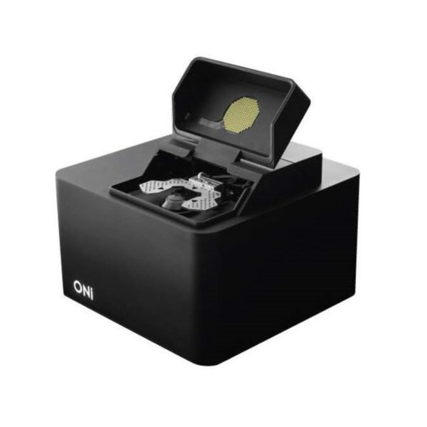
A microscope with a smaller footprint than an A4 sheet of paper and a desktop light engine with powerful lasers - Robust
Super-resolution on your benchtop or biosafety cabinet, no need for an optical table or dark room - Easy to use
Quick start for imaging, no alignment or calibrations required and real time data build up - Flexible
Different imaging & illumination modes for either fixed and live cell imaging, and programmable workflows
FAQs
More Products
At ONI, our mission is to accelerate human discovery and combat disease by enabling everyone to see and understand the…


