Showing all 8 results

  • Bruker – Confocal Raman Microscope – SENTERRA II

    Raman imaging is an incredibly effective tool to create detailed chemical images based on a sample’s Raman spectra. In these images, every pixel is composed of a complete Raman spectrum. By interpretation of this spectral data, a false color ima...

    Raman imaging is an incredibly effective tool to create detailed chemical images based on a sample’s Raman spectra. In these images, every pixel is composed of a complete Raman spectrum. By interpretation of this spectral data, a false color image can be rendered to emphasize and characterize the sample’s properties like chemical structure or composition. The SENTERRA II features comprehensive Raman imaging as well as mapping capabilities, to analyze and generate chemical images. Meanwhile, the intelligent software supports you when needed and saves a time consuming and overcomplex interface for setting parameters. Our data acquisition was designed to be as efficient as possible and in combination with the precise sample stage Raman images are produced in a matter of seconds.
  • Bruker – FT-IR Spectrometer – LUMOS II

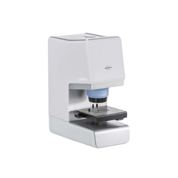
    The LUMOS II takes FTIR microscopy to the next level by making it faster, easier, more accurate and reliable. We accomplished this by tailoring its software and user interface specifically to the user. Beginners get perfect results in no time, whi...

    The LUMOS II takes FTIR microscopy to the next level by making it faster, easier, more accurate and reliable. We accomplished this by tailoring its software and user interface specifically to the user. Beginners get perfect results in no time, while experts are given almost unlimited possibilities. The LUMOS II offers exclusive FPA imaging technology, that naturally exceeds the speed and spatial resolution of line array and single-point measurements. Its applicability is enormous, the spectral data obtained always of the highest quality and the measurement times are very short.

  • Bruker – S2 POLAR – The EDXRF for the Petrochemical Industry

    The S2 POLAR masters all requirements for S analysis from diesel to crude oils. This also includes continuous process control of higher sulfur levels in refineries as well as norm-compliant Ultra-Low Sulfur (ULS) analysis of fin...

    The S2 POLAR masters all requirements for S analysis from diesel to crude oils. This also includes continuous process control of higher sulfur levels in refineries as well as norm-compliant Ultra-Low Sulfur (ULS) analysis of final products.

    The S2 POLAR complies with the international S norms such as ASTM D7220, D4294, and ISO 13032.

    With its capability to measure multiple elements simultaneously, including important elements in process control such as Ni, V, Fe and CI, the S2 POLAR is well suited for the analytical demands of refineries in one single instrument.

    The S2 POLAR is also well suited for the norm-compliant analysis of additives in lubricating oils according to ASTM D6481 and ASTM D7751 that includes the elements Mg, P, S, Cl, Ca, Zn, and Mo. Furthermore this also includes the analysis of elements in edible oils or palm oils.

  • Bruker – S2 PUMA Series 2 – High-End, Versatile Benchtop EDXRF

    Fast – Next generation SDD and 50 Watt Power

    A high-power (50 W), long-life-time X-ray tube combined with closely coupled optics and the new HighSenseâ„¢ detectors is the basis for unrivalled EDXR...

    Fast – Next generation SDD and 50 Watt Power

    A high-power (50 W), long-life-time X-ray tube combined with closely coupled optics and the new HighSenseâ„¢ detectors is the basis for unrivalled EDXRF performance.

    Intuitive – SPECTRA.ELEMENTS

    Next generation software based on state-of-the-art programming combines functionality with ease-of-use for a smooth and worry-free operation.

    Versatile – Optimal configuration for every need

    Depending on sample type and desired throughput, the versions Single, XY Autochanger, Carousel, Automation, and Mapping-Stage cover all possible scenarios.

  • Bruker – X-ray Micro-CT – SKYSCAN 1272

    High-Resolution Micro-CT

    Exceptional Performance Beyond Researcher’s Imaginations

    The system can nondestructively visualize up to 209 Megapixel (14450×14450 pixels) virtual slices through objects, more than 2600 such slices after a single scan using newly developed 16Mp and 11Mp X-ray detectors in up to three offset positions. Due to phase-contrast enhancement, object details as small as 0.35um can be detected.

    The innovative flexible acquisition geometry of the SKYSCAN 1272 scanner is particularly advantageous over intermediate resolution levels, where scans are several times faster (to obtain the same or better image quality) compared to micro-CT systems with a fixed source-detector design.

    For any sample, which can be up to 75mm in diameter, the system can automatically optimize x-ray energy and energy filtering using a new maintenance-free X-ray source and automatic 6-position filter changer. Large format imaging has been supported by world’s fastest hierarchical (InstaRecon®) and multithreaded GPU-accelerated 3D-reconstruction and realistic visualization by surface and volume rendering.


    The system includes a built-in precision micropositioning stage and can scan samples during compression, tension, cooling or heating. An optional 16-position automatic sample changer allows the adding / removing / replacing of samples without interrupting a live scanning process (see how it works). Automatic magnification adjustment and scanning protocol optimization is available for individual samples.

    At the end of the scan the system can send you an e-mail with a direct link to the scanning results, which can be opened across a network. Reconstruction results can be sent to iPad/iPhone/Android devices. Special software for mobile platform performs volume rendering and virtual cut of objects with touch screen gesture controls. The full range of the software for 2D / 3D quantitative analysis and for realistic 3D visualization is also supplied with the scanner.

    Supplied advanced workstation contains two Intel-XEON eight core processors, 128GB memory,  12TB disk space and two 24″ UltraSharp LCD monitors.

  • Bruker – X-ray Micro-CT – SKYSCAN 1273

    High-Capacity 3D X-ray Microscopy

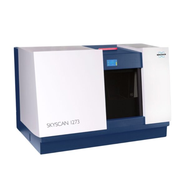
    The SKYSCAN 1273 is Bruker’s latest benchtop 3D X-ray microscope based on micro computed tomography (Micro-CT) technology. Samples with up to 500 mm length, 300 mm diameter, and a max...

    High-Capacity 3D X-ray Microscopy

    The SKYSCAN 1273 is Bruker’s latest benchtop 3D X-ray microscope based on micro computed tomography (Micro-CT) technology. Samples with up to 500 mm length, 300 mm diameter, and a maximum weight of 20 kg can be accommodated, a new standard for non-destructive testing (NDT) with benchtop instruments. Best-in-class components turn the SKYSCAN 1273 into a real power pack. The combination of a high-energy X-ray source running at high power and a large format flat-panel detector with ultimate sensitivity and speed provides excellent image quality in just a few seconds.

    The comprehensive software for straightforward data collection, advanced image analysis, and powerful visualization makes the SKYSCAN 1273 an easy to use 3D X-ray microscope.

    The SKYSCAN 1273 benchtop 3D X-ray microscope requires minimum lab space, is easy to start running, and virtually maintenance-free. Consequently, the SKYSCAN 1273 offers high system uptime with low cost of ownership.

  • Bruker – X-ray Micro-CT – SKYSCAN 1275

    Fast, Automated, Desk-top X-Ray Microtomograph

    Fast, Easy to Use and with no Compromise on Image Quality – the New Bruker SkyScan 1275 delivers a Winning Combination for Research and Industrial Markets

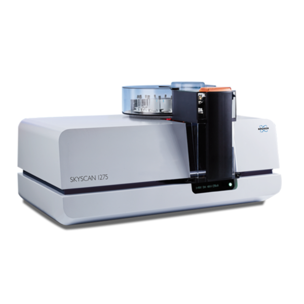
    The SkyScan1275 is specially designed for fast scanning using new advances in the technology of X-ray sources and efficient flat-panel detectors. Shortening the distance between source and detector and very fast camera readout opens the possibility for reducing scan time down to a few minutes without compromising image quality. Current developments in fast reconstruction accelerated by graphics cards give an additional gain in performance and speed.

    Fast realistic visualization of results by volume rendering enables fabulous imaging of internal object’s microstructure with power to reveal all internal details by virtual cut or virtual flight around and inside the objects. Fast scanning with high quality results is crucial for scientific research and industrial applications such as quality control or production process monitoring. The SkyScan1275 provides a high level of automation. Simple push of a button on the front of the system starts an auto-sequence of a fast scan followed by reconstruction and volume rendering executed during scanning of the next sample.

  • Bruker – X-ray Micro-CT – SKYSCAN 2214

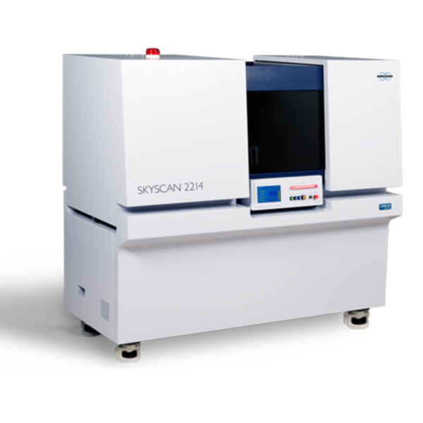
    The SKYSCAN 2214 is the latest nanotomograph by Bruker, the pioneer of microCT technology. The SKYSCAN 2214 offers X-ray microscopy at extreme resolutions with an unparalleled user experience. The latest technologies in each of the components make...

    The SKYSCAN 2214 is the latest nanotomograph by Bruker, the pioneer of microCT technology. The SKYSCAN 2214 offers X-ray microscopy at extreme resolutions with an unparalleled user experience. The latest technologies in each of the components make this the most complete and versatile system on the market today.

    The new Multiscale X-ray nanotomograph SKYSCAN 2214 covers the widest range of object sizes and spatial resolutions in one single instrument. It opens unique possibilities for 3D imaging and exact modeling of geological materials in oil and gas exploration, composite materials, Li batteries, fuel cells, electronic assemblies, as well as ex-vivo preclinical applications like in lung imaging or tumour vascularization.

    The instrument allows scanning and 3D non-destructive reconstruction of internal microstructure of objects as large as >300mm in diameter as well submicron resolution for small samples. The world’s fastest hierarchical 3D reconstruction software speeds-up imaging object’s internal microstructure 10-100 times compare to traditionally used algorithms or GPU-accelerated reconstruction.

    The system contains “open type” [pumped] X-Ray source with <0.5 microns spot size and diamond window. It can be equipped with one to four X-Ray detectors: flat-panel for large objects, 11Mp cooled CCD with wide field of view, 11Mp cooled CCD with medium field of view, 8Mp cooled CCD for highest spatial resolution. Automatically variable acquisition geometry and phase-contrast enhancement allow obtaining best possible quality in relatively short scanning time. The full range of Bruker-microCT software is supplied, including fast volumetric reconstruction, software for 2D/3D quantitative analysis and for realistic 3D visualization. Supplied software also includes volume rendering and easy creation of realistic movies with possibility for virtual flight around and inside the reconstructed object’s microstructure. Reconstruction results can be sent to iPad, iPhone or Android devices. Special software for mobile platforms performs volume rendering and virtual cut of objects with touch screen gesture controls.